TL;DR:
- Advanced dental technology integrates digital imaging, scanning, and manufacturing workflows to enhance treatment precision and patient comfort. It enables faster, more accurate restorations and implant placements through tools like CBCT, intraoral scanners, and AI-assisted planning, especially in private practices in London. Effective implementation relies on connected, well-trained protocols that ensure better outcomes and reduced appointments.
Advanced dental technology is defined as the integration of digital tools and computer-enabled workflows that allow dentists to diagnose, plan, and carry out cosmetic and restorative treatments with greater precision and less discomfort than traditional methods permit. For patients in Fulham and across London, this means treatments such as dental implants, crowns, veneers, and teeth straightening are now faster, more accurate, and more predictable than ever before. The core technologies driving this change include cone-beam computed tomography (CBCT), digital intraoral scanners, CAD/CAM fabrication systems, guided surgery, and artificial intelligence. Understanding how dental technology works as an integrated digital workflow, rather than a collection of individual devices, is the key insight that separates a well-equipped modern practice from one simply marketing itself as advanced.
What is advanced dental technology and why does it matter?
Advanced dental technology is best understood as a chain of connected digital stages: imaging, virtual planning, design, fabrication, and verification. Each stage feeds into the next, and the quality of the final result depends on how well those stages communicate with one another. A practice that uses a high-quality intraoral scanner but then relies on manual laboratory processes breaks that chain, and the benefits diminish accordingly.
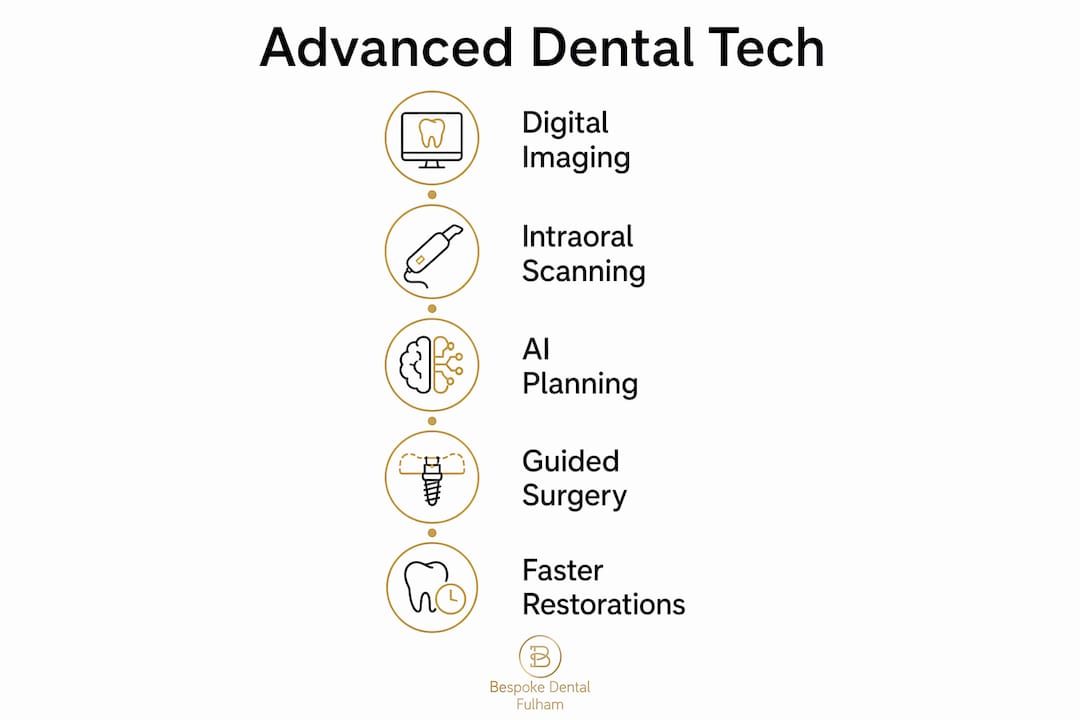
The technologies that make up this chain include:
- CBCT (cone-beam computed tomography): Three-dimensional X-ray imaging that maps bone volume, nerve pathways, and tooth roots with far greater detail than a standard two-dimensional radiograph.
- Digital intraoral scanners: Handheld devices that capture precise 3D models of your teeth and gums without the need for traditional impression putty.
- CAD/CAM systems: Computer-aided design and manufacturing tools that allow crowns, bridges, and veneers to be designed digitally and milled from ceramic or composite blocks, often within a single appointment.
- Guided surgery: Surgical templates produced from digital planning data that direct implant placement to within fractions of a millimetre.
- Artificial intelligence: Software that analyses imaging data to assist clinicians in identifying structures, planning implant positions, and flagging diagnostic concerns.
The practical result for you as a patient is fewer appointments, less guesswork, and restorations that fit more reliably from the outset. Practices in Fulham and Chelsea that have adopted closed-loop digital workflows report fewer remakes and adjustments because errors are caught at the planning stage rather than at the fitting stage.
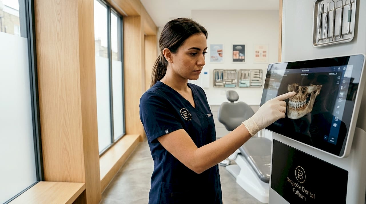
How do advanced imaging technologies improve diagnosis and treatment planning?
CBCT imaging is the foundation of accurate implant planning, and its advantages over traditional dental X-rays are substantial. A conventional two-dimensional radiograph shows height and width but not depth. CBCT produces a three-dimensional model of your jaw that reveals bone density, sinus proximity, nerve canal position, and the exact dimensions of the space available for an implant. This level of detail makes the difference between a procedure planned on assumptions and one planned on facts.
Radiation safety is a legitimate consideration, and responsible practices address it directly. Ultra-low-dose CBCT protocols reduce exposure to approximately 40.3 µSv compared with 89.0 µSv under standard-dose settings, without affecting the accuracy of surgical guides or patient-specific implant fit. This means you receive the diagnostic information your dentist needs at a meaningfully lower radiation dose. The principle guiding this approach is ALARA (as low as reasonably achievable), which professional guidance recommends as standard practice for all CBCT imaging.
Pro Tip: Ask your dentist whether they use the smallest field of view appropriate for your case. A targeted scan of a single implant site exposes you to far less radiation than a full-jaw scan, and dose optimisation is a mark of a clinician who takes your safety seriously.
CBCT data does not simply produce a picture. It feeds directly into surgical planning software, where your dentist can position implants virtually, check clearances, and generate a surgical guide before a single incision is made. For complex restorative cases in London, particularly those involving bone grafting or multiple implants, this planning stage is where the quality of your outcome is largely determined.
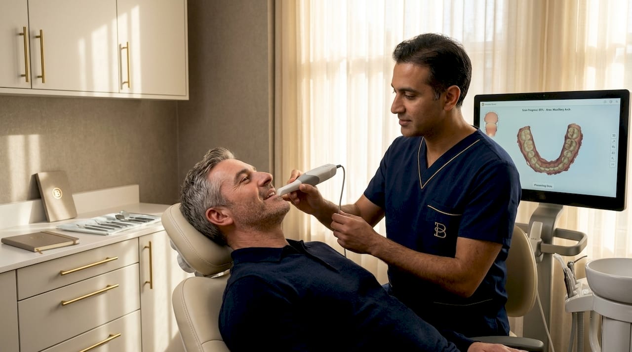
What are digital intraoral scanners and how do they improve dental restorations?
Digital intraoral scanners replace the traditional impression tray and putty with a small handheld wand that captures thousands of data points per second, producing an accurate 3D model of your teeth in real time. For patients who find conventional impressions uncomfortable or who have a sensitive gag reflex, this is a significant practical improvement. The digital model is transmitted directly to a dental laboratory or CAD/CAM milling unit, eliminating the distortion that can occur when physical impressions are poured, posted, and handled.
Scanner performance varies by device, and the differences matter in clinical practice. The table below summarises key findings from a 2026 clinical evaluation of three widely used scanners:
| Scanner | Full-arch scan time | Upper jaw trueness |
|---|---|---|
| Primescan 1 | 72.5 ± 3.8 seconds | Varies by region |
| Medit i700 | Longer than Primescan 1 | 100.3 ± 6.6 µm |
| Third device evaluated | Variable | Region-dependent |
The data shows that scanner accuracy varies by jaw, region of interest, and clinical scenario. Primescan 1 was the fastest for full-arch scanning, but speed alone does not determine which device is most appropriate for a given case. A dentist fitting a single crown on a lower molar requires different performance characteristics than one scanning a full arch for a bridge or implant-supported prosthesis.
Pro Tip: If you are having a crown or veneer made, ask whether your dentist uses a digital scanner rather than a traditional impression. Digital workflows reduce the margin for error and typically produce restorations that require less adjustment at the fitting appointment.
One area where intraoral scanners still face limitations is in scanning large edentulous (toothless) areas. Soft tissue lacks the fixed reference points that teeth provide, which can reduce accuracy. In these cases, experienced clinicians combine scanner data with CBCT imaging to compensate, rather than relying on a single data source.
How is artificial intelligence transforming dental implant planning?
AI in dentistry currently functions as a decision-support tool rather than an autonomous system, and that distinction matters. The most established application is the analysis of CBCT imaging data, where AI software segments bone, identifies teeth, maps nerve canals, and flags anatomical structures that require attention during implant planning. This process, which previously took a trained clinician considerable time, can now be completed in minutes.
The evidence for AI accuracy in this context is strong. AI systems analysing CBCT data achieve 92 to 99.7% accuracy in detecting teeth and edentulous regions, and implant planning success rates improve from 78% with traditional methods to 92% when AI-assisted planning is used. These are not marginal gains. For a patient undergoing a complex implant procedure, the difference between a 78% and 92% success rate in planning accuracy is clinically meaningful.
Current limitations are worth understanding:
- AI models trained on one imaging system may not perform equally well on data from a different scanner or CBCT unit.
- Silent errors can occur if imaging data does not meet the quality standards the AI model expects, making robust clinical validation a non-negotiable requirement.
- Ethical oversight and governance frameworks for AI in dentistry are still developing, and no AI system should be used without a qualified clinician reviewing and approving its outputs.
The future of AI in dentistry points towards earlier detection of decay, more accurate bone loss assessment in periodontal disease, and fully automated prosthetic design. For now, its greatest value lies in making implant planning faster, more consistent, and less dependent on individual clinician experience alone.
What practical benefits do patients in Fulham and London experience?
Patients attending private dental practices in Fulham, Parsons Green, Hammersmith, and Chelsea who benefit from these technologies typically notice the following differences compared with traditional dental care:
- Fewer appointments. Digital workflows compress the number of visits required. A crown that once required two or three appointments, including a temporary fitting, can often be completed in one session using CAD/CAM milling.
- Better-fitting restorations. Guided digital workflows reduce the accumulation of small errors across planning, design, and fabrication stages, producing implants, crowns, and veneers that seat more accurately from the first fitting.
- Less invasive procedures. Guided implant surgery, planned from CBCT data, allows dentists to place implants through smaller incisions with greater confidence, reducing post-operative discomfort and healing time.
- Clearer communication. Digital models and 3D visualisations allow your dentist to show you exactly what is planned before treatment begins, which supports informed decision-making and realistic expectations.
- Higher-quality aesthetic outcomes. For cosmetic treatments such as veneers and composite bonding, digital smile design tools allow the final result to be previewed and refined before any tooth preparation takes place, supporting the kind of smile confidence that patients in London increasingly expect from premium private care.
The private dental context in Fulham is relevant here. NHS dental provision does not routinely fund CBCT imaging, digital intraoral scanning, or AI-assisted planning. Access to these tools is a distinguishing feature of private practices that have invested in them, and it is worth asking any prospective dentist directly which technologies they use and how those tools are integrated into their clinical workflow.
Key takeaways
Advanced dental technology delivers its greatest patient benefit when digital imaging, scanning, design, and fabrication are integrated into a single closed-loop workflow rather than used as isolated tools.
| Point | Details |
|---|---|
| Integrated digital workflow | Technology works best as a connected chain from imaging to fabrication, not as individual devices. |
| CBCT imaging precision | 3D imaging enables accurate implant planning and surgical guide creation with dose-optimised protocols. |
| Scanner selection matters | Accuracy varies by device, jaw, and clinical scenario; the right scanner depends on your specific case. |
| AI as clinical support | AI achieves over 90% accuracy in implant planning analysis but requires qualified clinician oversight. |
| Private care advantage | Advanced tools such as CBCT, intraoral scanning, and guided surgery are standard in quality private practices, not NHS provision. |
Why technology alone does not guarantee a great result
I have spent considerable time reviewing how digital dental technology is adopted across private practices in London, and the pattern I see most often is this: practices invest in impressive equipment but underinvest in the clinical protocols that make that equipment perform. A CBCT unit is only as useful as the clinician interpreting its output. An intraoral scanner produces accurate data only if the scanning technique is correct and the software workflow is properly configured end to end.
The closed-loop principle is the one I return to most often when advising patients. Small inconsistencies accumulate across digital workflow stages, and practices that control every step from scanning through to manufacturing verification consistently produce better-fitting restorations than those that mix digital and analogue processes. When you are evaluating a practice in Fulham or across London, the question to ask is not “do you use digital technology?” but “how does your digital workflow connect from scan to final restoration?”
I also think patients underestimate the importance of radiation governance. The fact that ultra-low-dose protocols maintain clinical adequacy for surgical planning is genuinely good news, but only if the practice is actually using them. Asking your dentist about their CBCT dose protocols is a reasonable and informed question, not an intrusive one. A confident, well-trained clinician will welcome it.
The future of dentistry in London is clearly digital, and the benefits of aesthetic dental treatments available to patients today would have been unrecognisable a decade ago. But technology serves the clinician, not the other way around. The best outcomes come from practices where skilled dentists use these tools with genuine clinical rigour, not simply as a marketing differentiator.
— Amit
Experience advanced dental care at Bespokedentalfulham
Bespokedentalfulham uses 3D CBCT imaging, digital intraoral scanning, and guided surgical workflows as standard across its cosmetic and restorative treatments in Fulham, London. Whether you are considering dental implants in Fulham or exploring cosmetic options such as veneers, composite bonding, or Invisalign, every treatment plan begins with precise digital assessment rather than guesswork. The practice operates at Harley Street standard, combining digital smile design with personalised care in a discreet, comfortable environment suited to patients across SW6, Chelsea, Parsons Green, and Hammersmith.
To explore how these technologies can support your treatment, visit the cosmetic dentistry page or contact Bespokedentalfulham directly to arrange a consultation.
FAQ
What is advanced dental technology in simple terms?
Advanced dental technology refers to digital tools such as 3D imaging, intraoral scanners, and computer-aided design systems that allow dentists to plan and carry out treatments with greater accuracy and less discomfort than traditional methods.
Is CBCT imaging safe for dental patients?
CBCT imaging is safe when used appropriately. Ultra-low-dose protocols reduce radiation exposure to around 40.3 µSv, and professional guidance recommends using the smallest field of view necessary for each case to keep exposure as low as possible.
How accurate are digital intraoral scanners compared with traditional impressions?
Digital intraoral scanners are highly accurate for most clinical scenarios, though performance varies by device and jaw region. Primescan 1 completes a full-arch scan in approximately 72.5 seconds, and accuracy is generally superior to traditional impressions for fixed prosthetic work.
Can AI replace a dentist in implant planning?
AI cannot replace a dentist. It functions as a decision-support tool that analyses CBCT data and assists with implant positioning, achieving over 90% accuracy in detecting anatomical structures, but all outputs require review and approval by a qualified clinician.
Do I need to go private to access advanced dental technology in London?
Advanced tools such as CBCT imaging, digital intraoral scanning, and guided surgery are not routinely available through NHS dental provision. Private practices in Fulham and across London that have invested in these technologies offer patients access to more precise and efficient treatment options.